Use microscopy-led records to compare bacterial morphology, then cross into biological structural color without collapsing scale, taxonomy or mechanism.
Media boundaryVisualization — source-backed explainer, not documentary media. This local SVG remains explanatory media, not documentary source media.
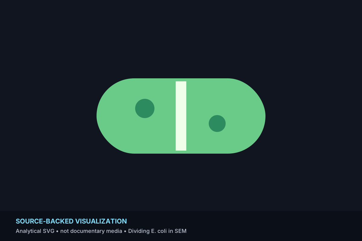
A scanning electron micrograph makes the dividing wall of Escherichia coli visible at a scale far below unaided sight.
Source-backed record material
CDC PHIL identifies the source as a digitally colorized scanning electron micrograph at 21,674× showing a dividing E. coli cell wall. The visible texture and form are mediated by electron microscopy and colorization.
Media boundaryVisualization — source-backed explainer, not documentary media. This local SVG remains explanatory media, not documentary source media.
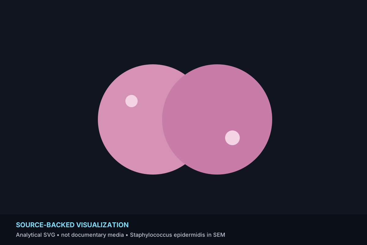
Scanning electron microscopy reveals paired spherical Staphylococcus epidermidis cells as discrete three-dimensional forms.
Source-backed record material
CDC PHIL identifies the source as a scanning electron micrograph of Staphylococcus epidermidis. The microscopy converts surface structure at microbial scale into an image readable at human scale.
Media boundaryVisualization — source-backed explainer, not documentary media. This local SVG remains explanatory media, not documentary source media.
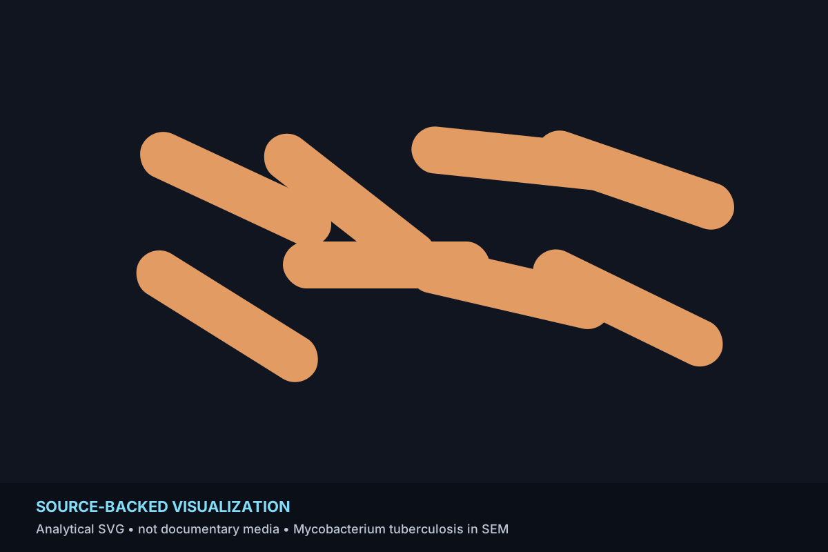
A digitally colorized SEM turns slender Mycobacterium tuberculosis cells into a highly legible microscopic field.
Source-backed record material
CDC PHIL describes a digitally colorized SEM at 15,549× and gives typical cell dimensions of roughly 2–4 micrometers long and 0.2–0.5 micrometers wide, explaining the elongated rod-like forms.
Media boundaryVisualization — source-backed explainer, not documentary media. This local SVG remains explanatory media, not documentary source media.
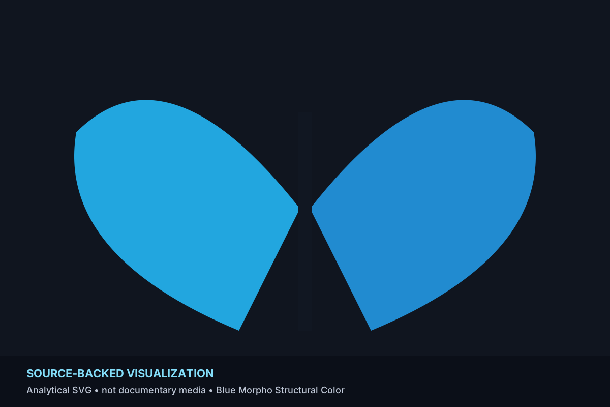
A blue morpho butterfly produces brilliant blue through microscopic wing-scale structure rather than blue pigment alone.
Source-backed record material
Smithsonian educational material explains that microscopic grooves and layers in morpho wing scales selectively reinforce blue reflected light while other wavelengths cancel, producing intense structural color.
The trail leaves microbial morphology for a biological microstructure/color record. This final bridge is editorial and does not create a biological or mechanistic relationship.
No normalized relationship record is created by this transition.
Connected discovery
Continue through reviewed visual records
These parent recommendations are bounded and non-sponsored. Curated journey order does not change canonical relationship evidence.