
Visual Entry · evidence-aware deep view
Mycobacterium tuberculosis in SEM
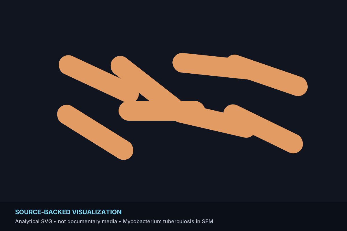
A digitally colorized SEM turns slender Mycobacterium tuberculosis cells into a highly legible microscopic field.
Visual literacy · read before concluding
Four questions for this image
Awe is the invitation. Interpretation starts by separating the subject, scale, representation, mechanism, and evidence.
1 · What am I looking at?
A digitally colorized SEM turns slender Mycobacterium tuberculosis cells into a highly legible microscopic field.
Displayed: visualization
2 · At what scale?
Microscopic. Below unaided sight; instrument-mediated detail. Reviewed record anchor: roughly 2–4 micrometers long and 0.2–0.5 micrometers wide.
3 · How was it represented?
Displayed media: Deterministic ICU-authored vector explanatory visualization.
Reviewed source method: Scanning electron microscopy + digital colorization
The reviewed claim explicitly identifies SEM and digital colorization.
4 · What creates the visible effect?
source backed: CDC PHIL describes a digitally colorized SEM at 15,549× and gives typical cell dimensions of roughly 2–4 micrometers long and 0.2–0.5 micrometers wide, explaining the elongated rod-like forms.
Current visual-property tags: geometry, texture.
Explanation boundary
Why it looks this way
Only source-backed claims can carry the mechanism/explanation role here. Curated or unknown material stays visibly separate.
CDC PHIL describes a digitally colorized SEM at 15,549× and gives typical cell dimensions of roughly 2–4 micrometers long and 0.2–0.5 micrometers wide, explaining the elongated rod-like forms.
Provenance & evidence
Know what supports the entry.
Source pages support subject identity and claims. The displayed SVG has its own representation and rights record.
It describes the relationship between a factual statement and reviewed evidence. It does not mean the SVG shown on this page is a source photograph, museum image or scientific plate.
Mycobacterium tuberculosis SEM
- Canonical locator
- https://wwwn.cdc.gov/phil/details.aspx?pid=9997
- Source date
- Not specified in record
- Creator / author
- Janice Haney Carr
- Rights note
- CDC PHIL record identifies the source image as Public Domain; factual/provenance use only here.
Runtime-media boundary
Documentary source pages reviewed. Published runtime media is an original explanatory SVG visualization, not a copied source image.
- reviewed_usable · Original deterministic SVG explanatory visualizations authored for BVU004. These files are ICU-owned presentation media and are visibly labeled as visualizations, not documentary source images.
Displayed media SHA-256: d494e8f2f7dddc4f32642b4fdd6e8fc528c96d0b0c27a2817910dcc5edde5706
Documentary-media review
This entry keeps its reviewed institutional source pages as provenance/factual references. The current media audit did not promote documentary bytes because explicit reusable rights and same-origin original-byte localization were not both cleared in this bounded pass.
- Runtime image
- Existing same-origin explanatory SVG visualization
- Documentary runtime
- Not published
- Remote hotlink
- Not used
Typed connections
Follow the relationship without upgrading its evidence.
Directionality and evidence class are rendered from the canonical relationship records.
Helicobacter bilis in SEM
Both are microscopy-led studies of bacterial morphology; no taxonomic equivalence is implied.
Curated interpretation: a deliberate discovery connection, not proof of shared cause, influence or mechanism.
Staphylococcus epidermidis in SEM
Both use electron microscopy to compare microbial geometries at scales below unaided sight.
Curated interpretation: a deliberate discovery connection, not proof of shared cause, influence or mechanism.
Collections & Atlas
Return through a useful visual lens.
Collection membership is editorial discovery context, not proof of shared mechanism or origin.
Microscopic Forms
Structures whose decisive details emerge only through microscopy or microscopic explanation.
BVU004 editorial collection. Membership indicates a useful discovery grouping, not shared origin, influence or mechanism.
Curated journeys
See where editors place this entry next.
These paths are editorial navigation. They do not alter this entry’s claim, provenance or relationship evidence state.