Skip to Breathtaking Visuals Universe content
Infinite Concept Universeresearch · make · build · play
Navigate the universe

Choose a direction.

Plan 5 treats the original eight worlds as historical origins, not mandatory top-level navigation.

Curated Collection · editorial lens

Microscopic Forms

Structures whose decisive details emerge only through microscopy or microscopic explanation.

Compare forms that become legible through microscopy or microscopic explanation without treating similar shapes as taxonomic or mechanistic equivalence.

Exact membership

Why these records sit together

Every card links back to the full Visual Entry, where source, provenance, rights and relationship evidence remain authoritative.

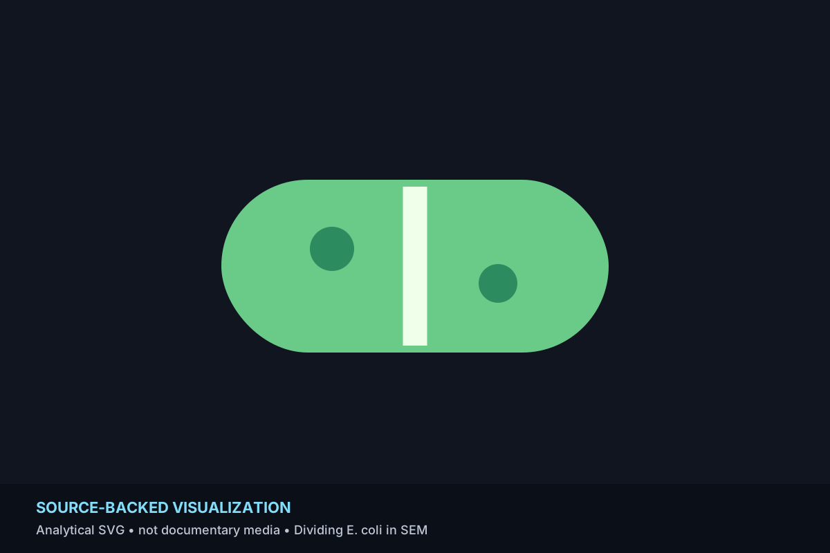
Simplified explanatory visualization for Dividing E. coli in SEM; not documentary source media.

Source Backed

Dividing E. coli in SEM

A scanning electron micrograph makes the dividing wall of Escherichia coli visible at a scale far below unaided sight.

Why it belongs — editorialEditorial membership is based on the published BVU metadata for this entry: Microscopic, Scientific Imaging, Biology; visual lens: Texture, Geometry. Membership is a discovery grouping, not a new claim about shared cause or origin.
Source-backed entry materialCDC PHIL identifies the source as a digitally colorized scanning electron micrograph at 21,674× showing a dividing E. coli cell wall. The visible texture and form are mediated by electron microscopy and colorization.
Simplified explanatory visualization for Staphylococcus epidermidis in SEM; not documentary source media.

Source Backed

Staphylococcus epidermidis in SEM

Scanning electron microscopy reveals paired spherical Staphylococcus epidermidis cells as discrete three-dimensional forms.

Why it belongs — editorialEditorial membership is based on the published BVU metadata for this entry: Microscopic, Scientific Imaging, Biology; visual lens: Geometry, Texture. Membership is a discovery grouping, not a new claim about shared cause or origin.
Source-backed entry materialCDC PHIL identifies the source as a scanning electron micrograph of Staphylococcus epidermidis. The microscopy converts surface structure at microbial scale into an image readable at human scale.
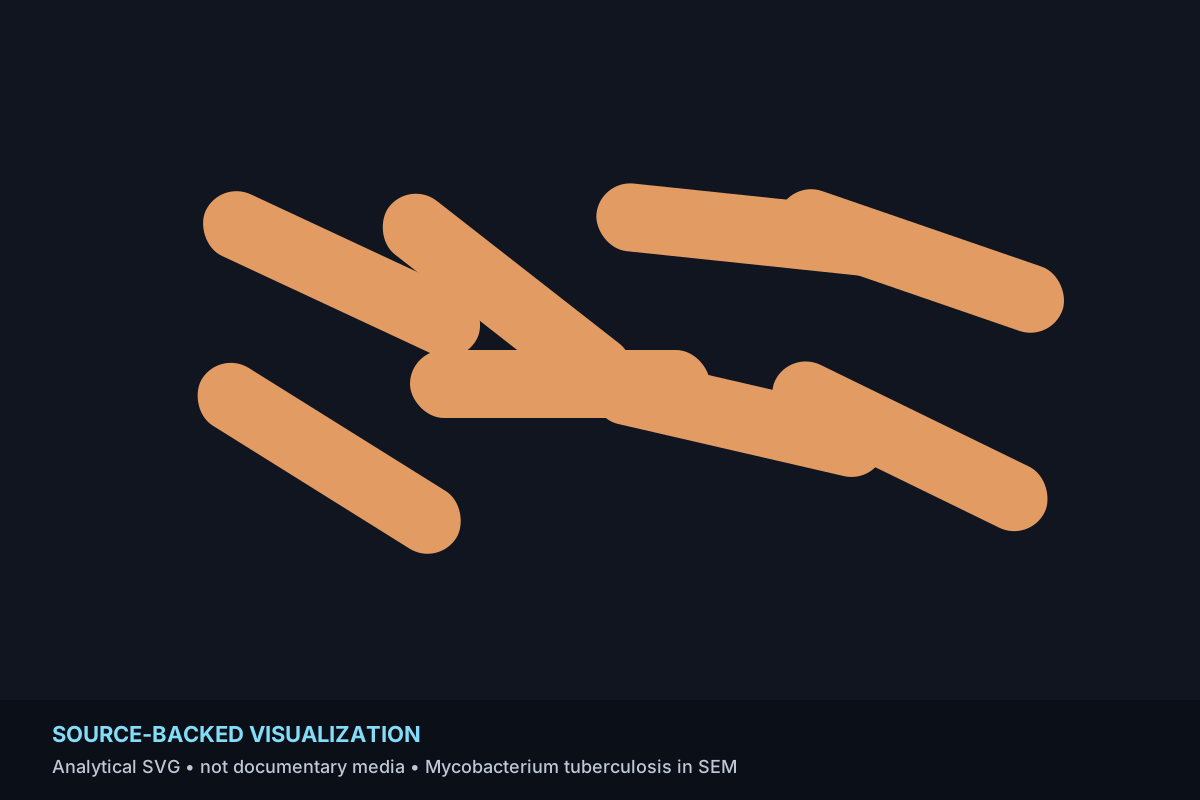
Simplified explanatory visualization for Mycobacterium tuberculosis in SEM; not documentary source media.

Source Backed

Mycobacterium tuberculosis in SEM

A digitally colorized SEM turns slender Mycobacterium tuberculosis cells into a highly legible microscopic field.

Why it belongs — editorialEditorial membership is based on the published BVU metadata for this entry: Microscopic, Scientific Imaging, Biology; visual lens: Geometry, Texture. Membership is a discovery grouping, not a new claim about shared cause or origin.
Source-backed entry materialCDC PHIL describes a digitally colorized SEM at 15,549× and gives typical cell dimensions of roughly 2–4 micrometers long and 0.2–0.5 micrometers wide, explaining the elongated rod-like forms.
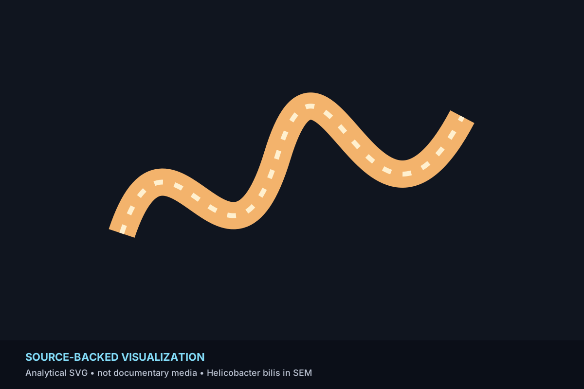
Simplified explanatory visualization for Helicobacter bilis in SEM; not documentary source media.

Source Backed

Helicobacter bilis in SEM

Electron microscopy reveals the spiral form, fibers and flagellar structures of Helicobacter bilis.

Why it belongs — editorialEditorial membership is based on the published BVU metadata for this entry: Microscopic, Scientific Imaging, Biology; visual lens: Geometry, Texture. Membership is a discovery grouping, not a new claim about shared cause or origin.
Source-backed entry materialCDC PHIL describes a 13,951× scanning electron micrograph showing spiral morphology, periplasmic fibers and bipolar tufts of sheathed flagella.
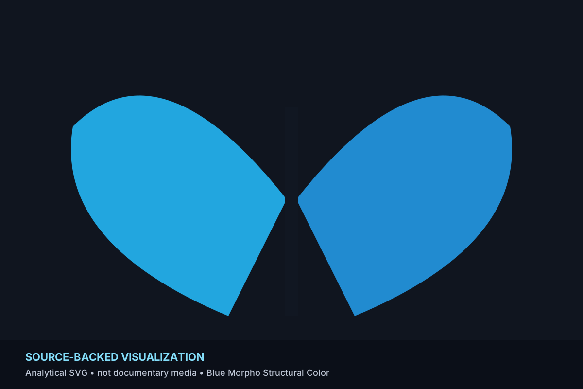
Simplified explanatory visualization for Blue Morpho Structural Color; not documentary source media.

Source Backed

Blue Morpho Structural Color

A blue morpho butterfly produces brilliant blue through microscopic wing-scale structure rather than blue pigment alone.

Why it belongs — editorialEditorial membership is based on the published BVU metadata for this entry: Biology, Light Color Optics, Extreme Scale; visual lens: Iridescence, Color. Membership is a discovery grouping, not a new claim about shared cause or origin.
Source-backed entry materialSmithsonian educational material explains that microscopic grooves and layers in morpho wing scales selectively reinforce blue reflected light while other wavelengths cancel, producing intense structural color.

Continue

Change the lens without changing the facts.